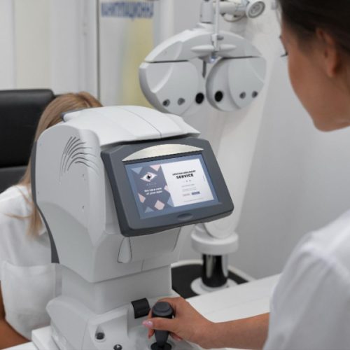
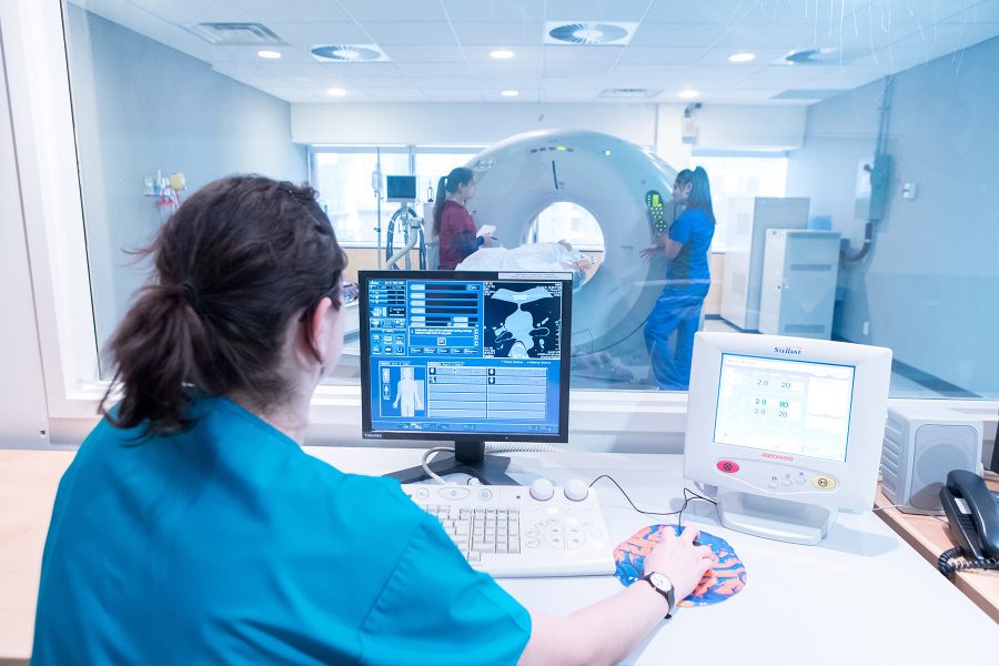
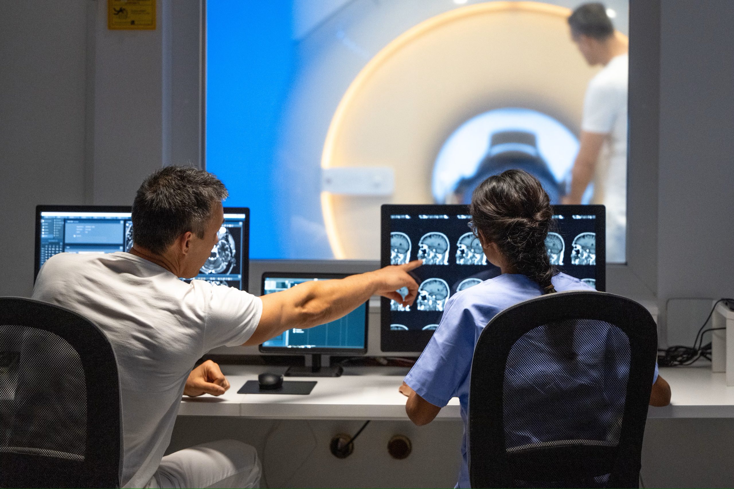
Medical professionals rely on crisp, clear imaging to make life-impacting decisions. In radiology, where even subtle contrast differences can indicate major conditions, display consistency becomes non-negotiable. This is where medical display calibration steps in—not just as a technical necessity but as a fundamental layer of diagnostic accuracy.
Display calibration ensures that grayscale medical images appear consistently across different monitors, regardless of the room, system, or user. Medical images stored in PACS systems are built from digital signals, and these need to be displayed faithfully.
The software PerfectLum, developed by QUBYX, plays a vital role in ensuring that medical monitors maintain conformity to calibration standards over time. QUBYX has earned its reputation for delivering highly reliable calibration tools that help healthcare systems meet stringent imaging standards.
How our eyes notice the difference between light and dark shades is mapped by the DICOM curve, which is a main principle used in calibration. It is not a linear relationship—the human eye is more sensitive to differences in dark tones than in bright ones.
If displays are calibrated according to DICOM standards, viewing images is similar to viewing film originals and makes sure everyone gets similar results when interpreting, regardless of the screen used.
One essential component in medical displays, especially in NY PDM, which stands for New York Primary Diagnostic Monitor, is the AMLCD (Active Matrix LCD) panel. These panels use a complex matrix of pixels that allow light from a backlight to pass through based on voltage inputs.
But not all pixels react the same way. Each one has its own voltage-to-luminance response curve, and these are mapped precisely during factory calibration. However, aging of the backlight or environmental changes can affect performance over time, making recalibration necessary.
To maintain DICOM calibration, there are two main methods used in the industry. One uses a sensor placed behind the display, directly measuring the backlight’s output. The other uses a tiny sensor on the front corner of the screen, capturing the luminance after it passes through the liquid crystal and filters.
Each approach comes with pros and cons. The back sensor provides more accurate and less noise-prone measurements, especially in the central viewing area. However, it cannot detect if the front display matrix fails.
On the other hand, the front sensor reflects the complete light path, catching issues in both the liquid crystal layers and the backlight, but may sacrifice accuracy due to its limited size and corner position.
Whichever method is used; calibration is not a one-time event. LCD monitors need initial setup and ongoing adjustments. Without calibration, visual consistency degrades, affecting diagnostic accuracy.
Healthcare facilities should prioritize displays that maintain their luminance and grayscale performance over time. Proper use of calibration tools such as PerfectLum means your diagnostic imaging will be both reliable and accurate for years.
It is not only about creating great-looking presentations but also about improving how we look after patients. For professionals making daily decisions from grayscale images, calibrated monitors are the unsung heroes behind every confident diagnosis.